EQUIPMENT
Dr. Sarafianos’ laboratory is fully equipped for state of the art structural, biochemical, virological, and molecular biology studies.
For protein chemistry and general molecular biology work:
The Sarafianos lab has three AKTA FPLC systems (Prime Plus, Purifier 100, Pure with sample pump, and Start systems)
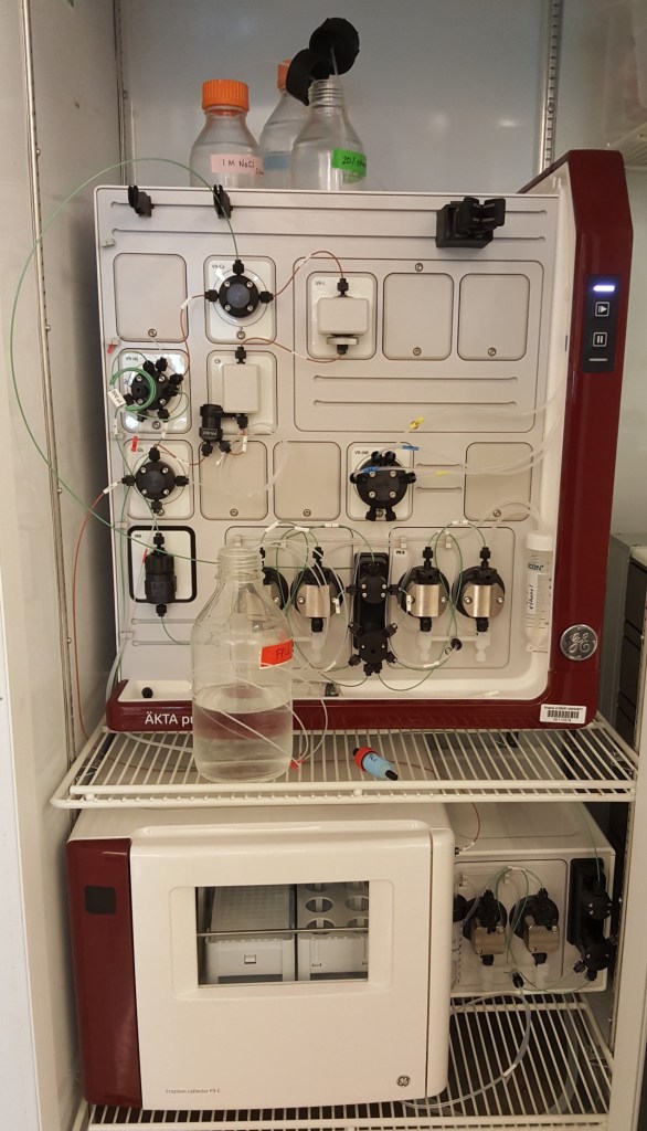
AKTA Pure
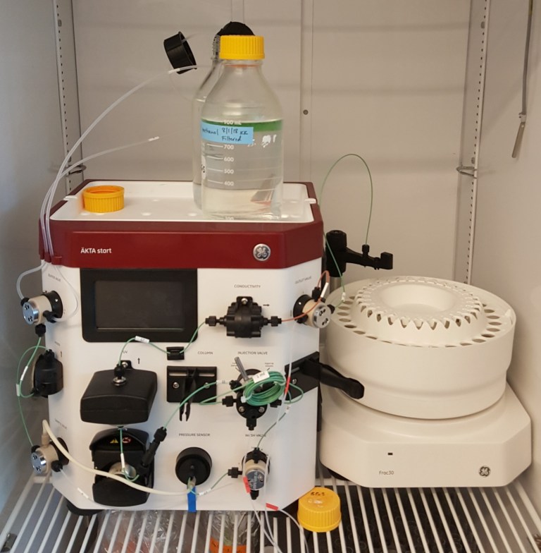
AKTA Start
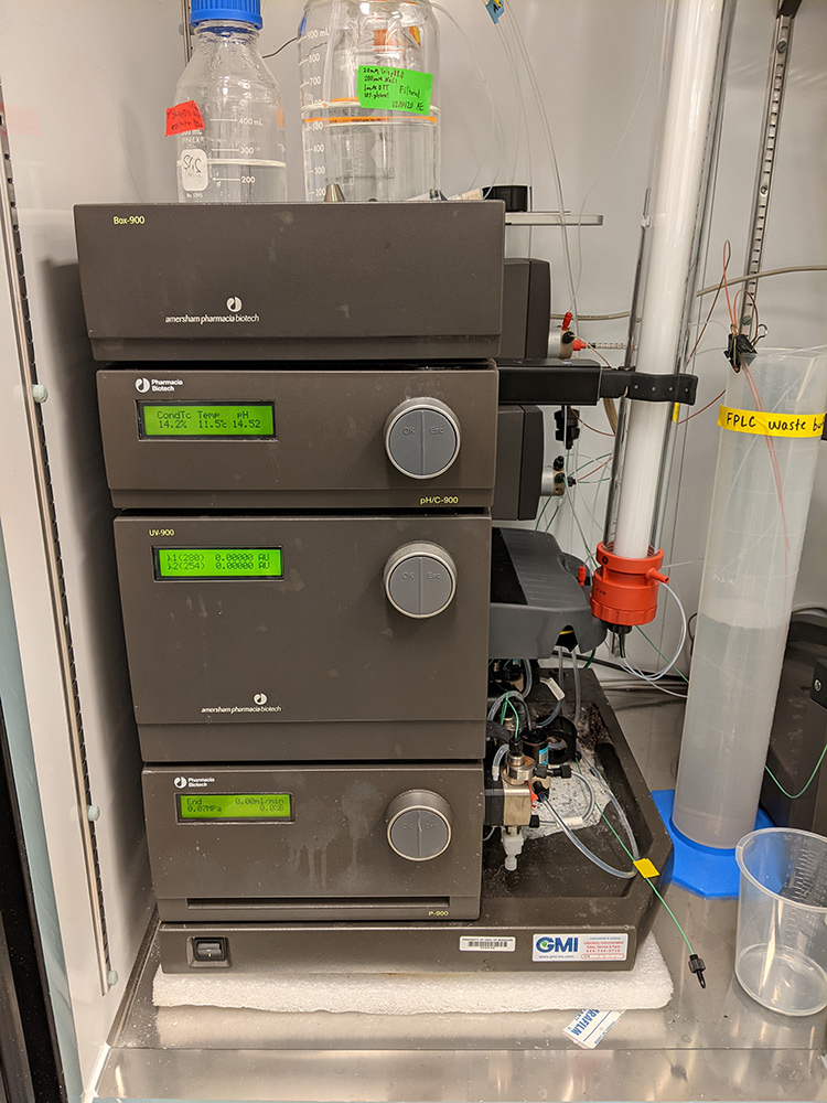
AKTA Purifier 100
For structural experiments:
The lab recently acquired a Wyatt Technologies DynaPro II 96-well dynamic light scattering (DLS) plate reader equipped with a temperature control unit for evaluation of protein aggregation state and molecular weight. This DLS instrument can also assess the stability of proteins and protein complexes for downstream crystallographic and cryo-EM structural studies. The lab acquired a Wyatt Technologies Dawn Helios II multi-angle light scattering (MALS) instrument coupled to an Agilent 1100 HPLC for performing size exclusion chromatography coupled with MALS (SEC-MALS) experiments (pictured right) for evaluating molecular weight and stability of proteins and large protein complexes for structural studies. We have access to a 4 °C cold room and own 12 °C and 18 °C incubators for growing crystals and 4 microscopes for viewing crystals. The lab owns an Art Robbins Instruments Crystal Gryphon “crystallization robot” and a CrysCam digital microscope that can record crystal growth in 96-well plates.
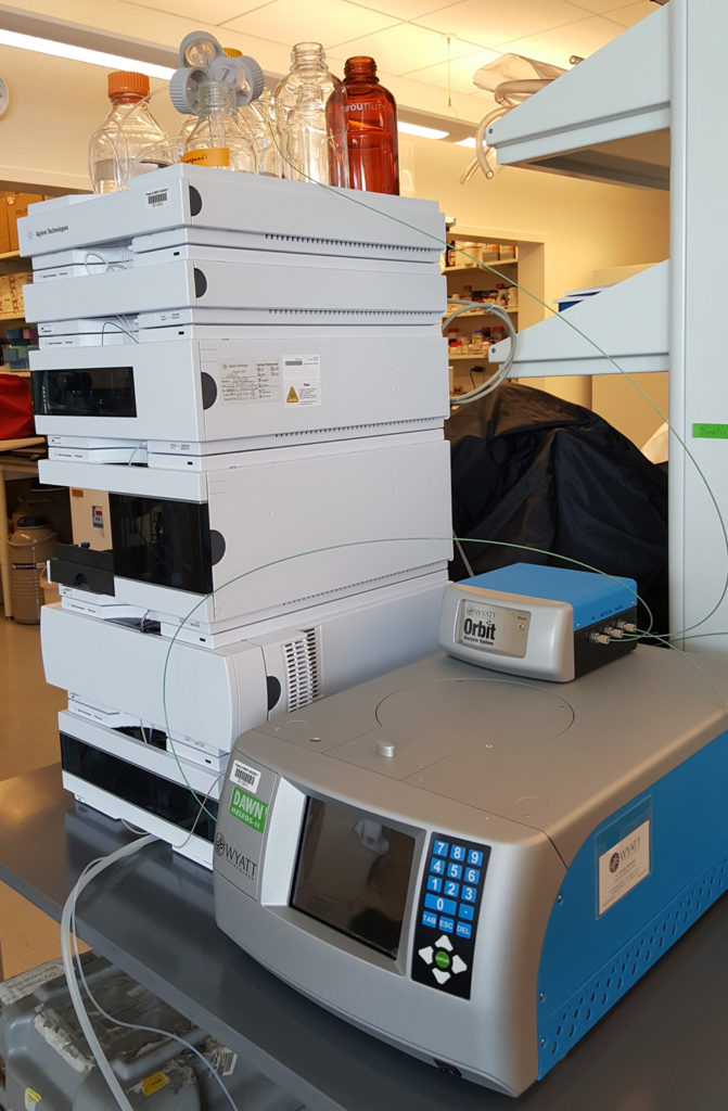
SEC-MALS
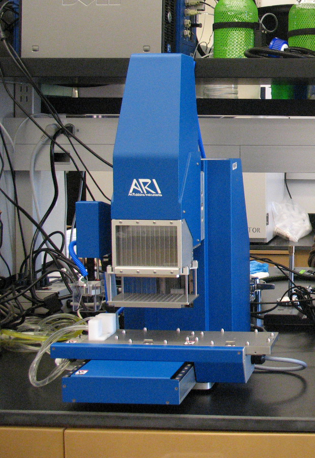
Crystal Gryphon
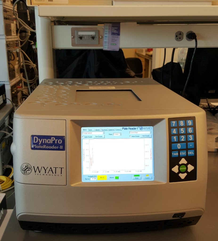
DynaPro II 96-well DLS
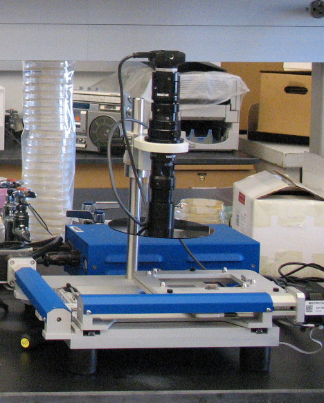
CrysCam
For additional screening of crystals, the lab has a JANSi UVEX-m brightfield/UV fluorescence and UV absorbance microscope to identify small protein crystals, for example in precipitate, that would otherwise be difficult to detect by visualization using only traditional brightfield microscopy. For screening crystals and collecting data, the lab has access to s X-ray Crystallography Center which houses a Rigaku XtaLAB Synergy-S high intensity dual-source microfocus X-ray diffractometer equipped with a HyPix-6000HE Hybrid Photon Counting (HPC) detector. The Sarafianos Lab also has an active general user proposal at the Advanced Photon Source (APS) at Argonne National Lab in Lemont, IL and has been collecting data at GM/CA CAT beamlines 23-ID-B and 23-ID-D, which have microbeam capabilities. Additionally, we are a member of the SER-CAT beamlines (22-ID and 22-BM) at APS
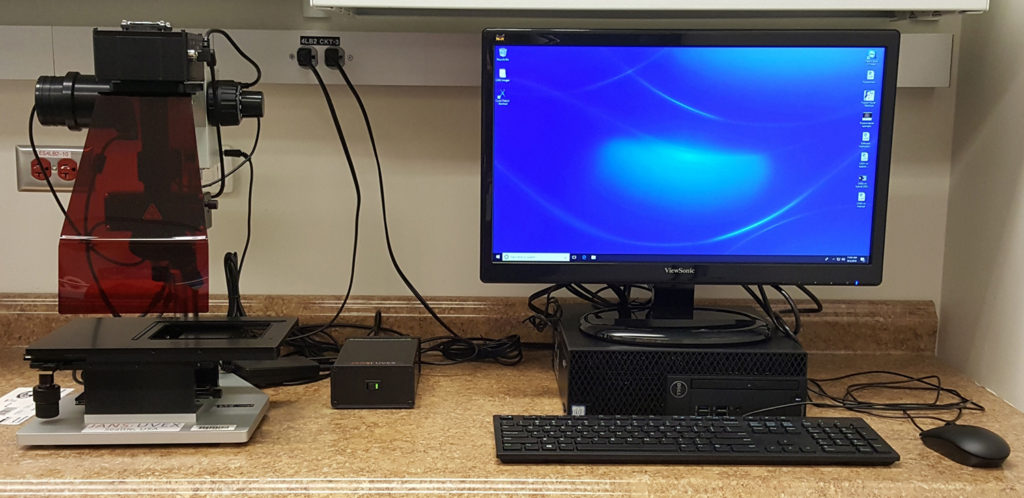
UVEX-m
The Sarafianos lab has state-of-the-art kinetics instrumentation for stopped-flow kinetics (Kintek SF-100). For evaluation of protein-protein, protein-nucleic acid, and protein-inhibitor interactions, the lab acquired a Monolith NT.115 thermophoresis instrument from NanoTemper Technologies. The Monolith NT.115 measures the thermophoresis of fluorescently labeled molecules, in any buffer or complex bioliquid, with low sample consumption. Using the Monolith NT.115, the binding affinities (Kd) of protein-protein, nucleic acid, or small molecule complexes can be determined. The Monolith NT.115 also provides information on sample aggregation. The Sarafianos lab also has a NanoTemper Tycho NT.6 which can analyze the quality of a protein sample in its native state in 3 minutes. Protein analysis can also be performed in a variety of buffers for assay development.
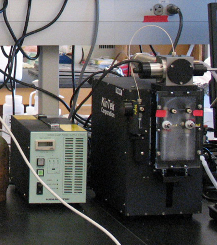
Stopped Flow
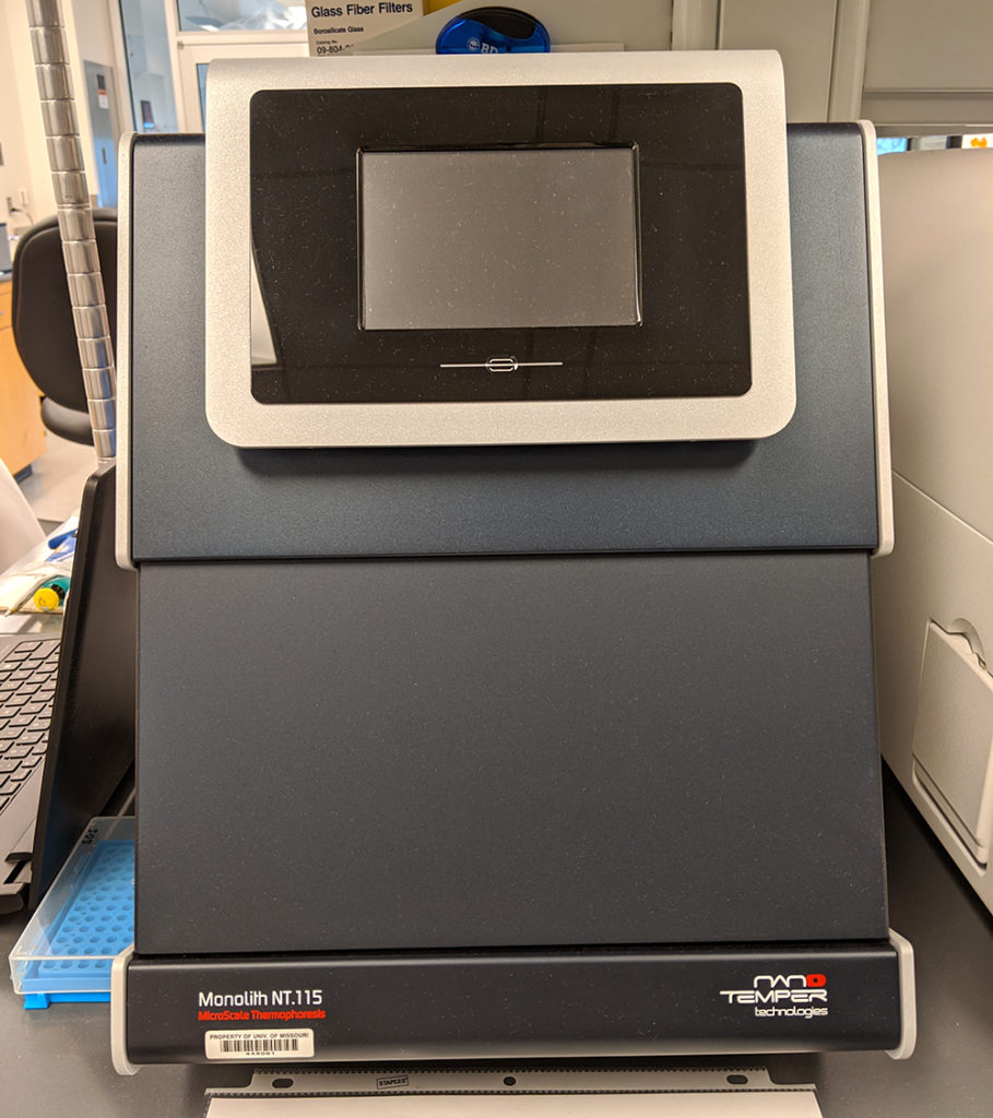
Monolith NT.115
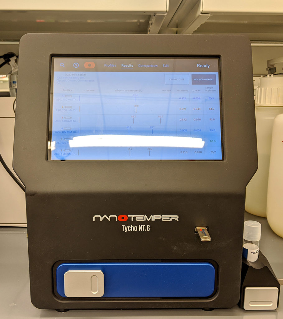
Tycho NT.6
The Sarafianos lab has a BioTek Synergy Neo2 high-throughput multimode plate reader for luminescence, fluorescence, and absorbance assays. The Sarafianos lab also has both a ThermoFisher Scientific QuantStudio 3 Real-Time PCR System and a Thermo Scientific PikoReal Real-Time PCR System for qPCR analyses and thermal shift assays to screen for ligands/proteins that bind target proteins. Protein melting curves are analyzed using the associated QuantStudio Design and Analysis or PikoReal software package. The Sarafianos lab also has a Typhoon FLA 9000, which is a versatile laser scanner used to image biochemistry gels, and a New Brunswick Innova 44R refrigerated shaking incubator for bacteria induction and protein expression.
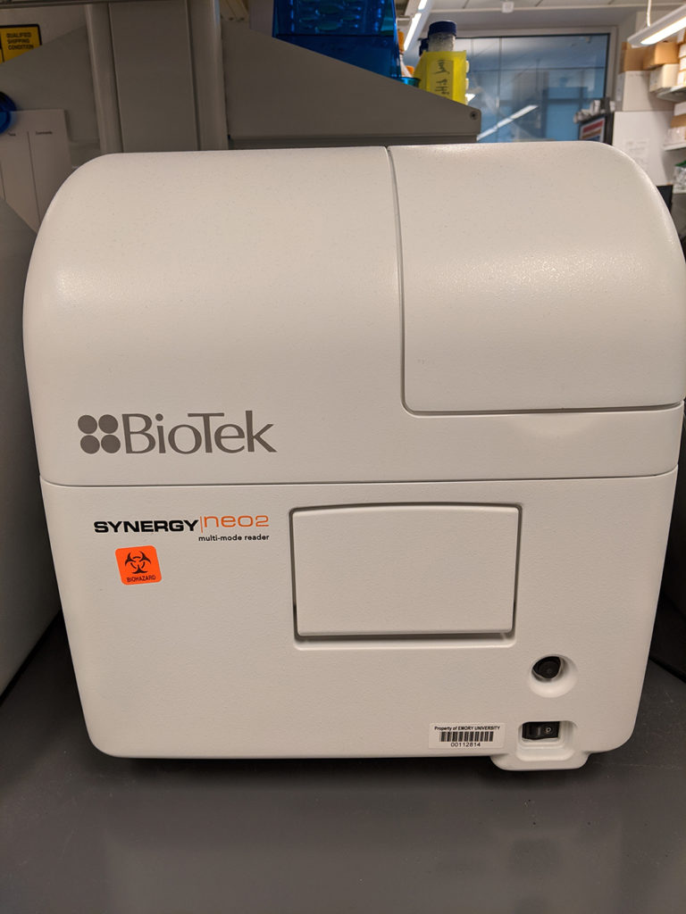
BioTek Synergy Neo2
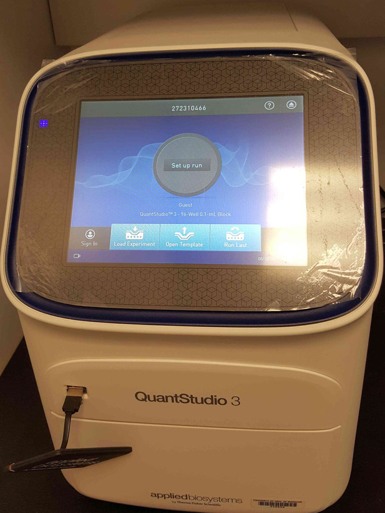
QuantStudio 3
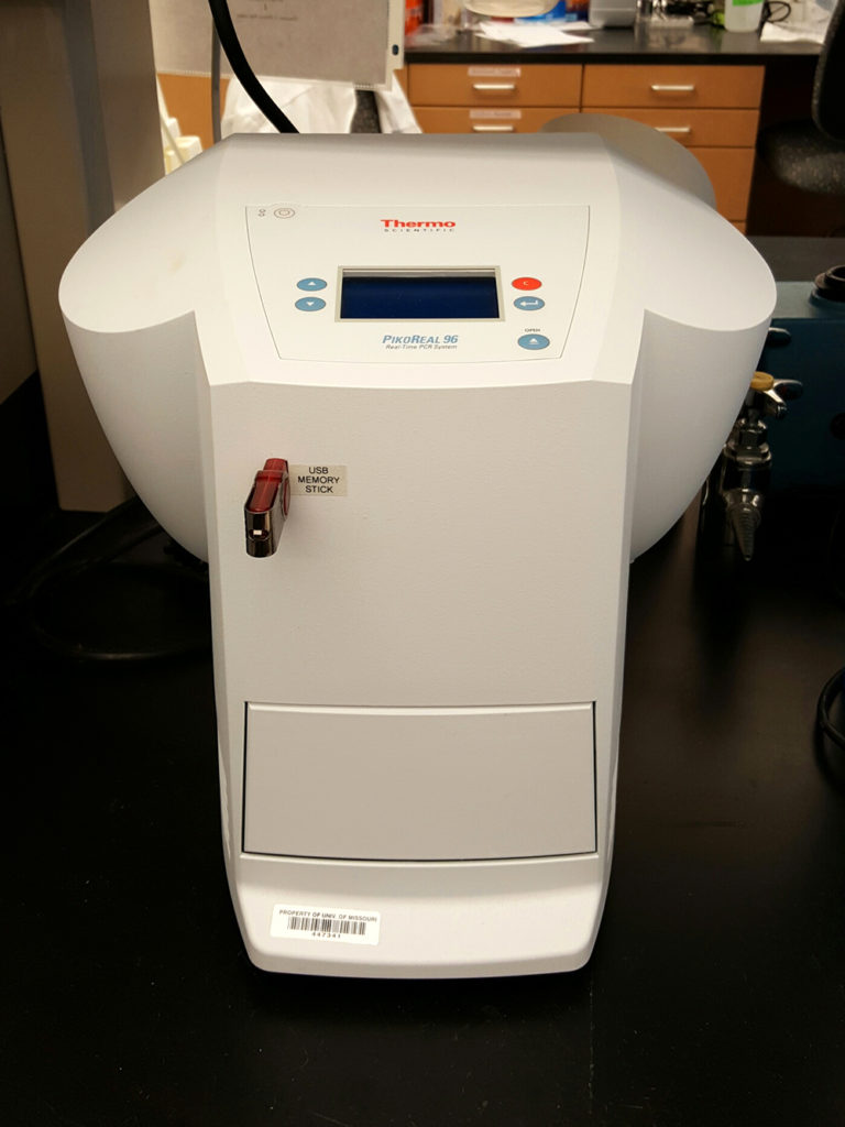
PikoReal 96
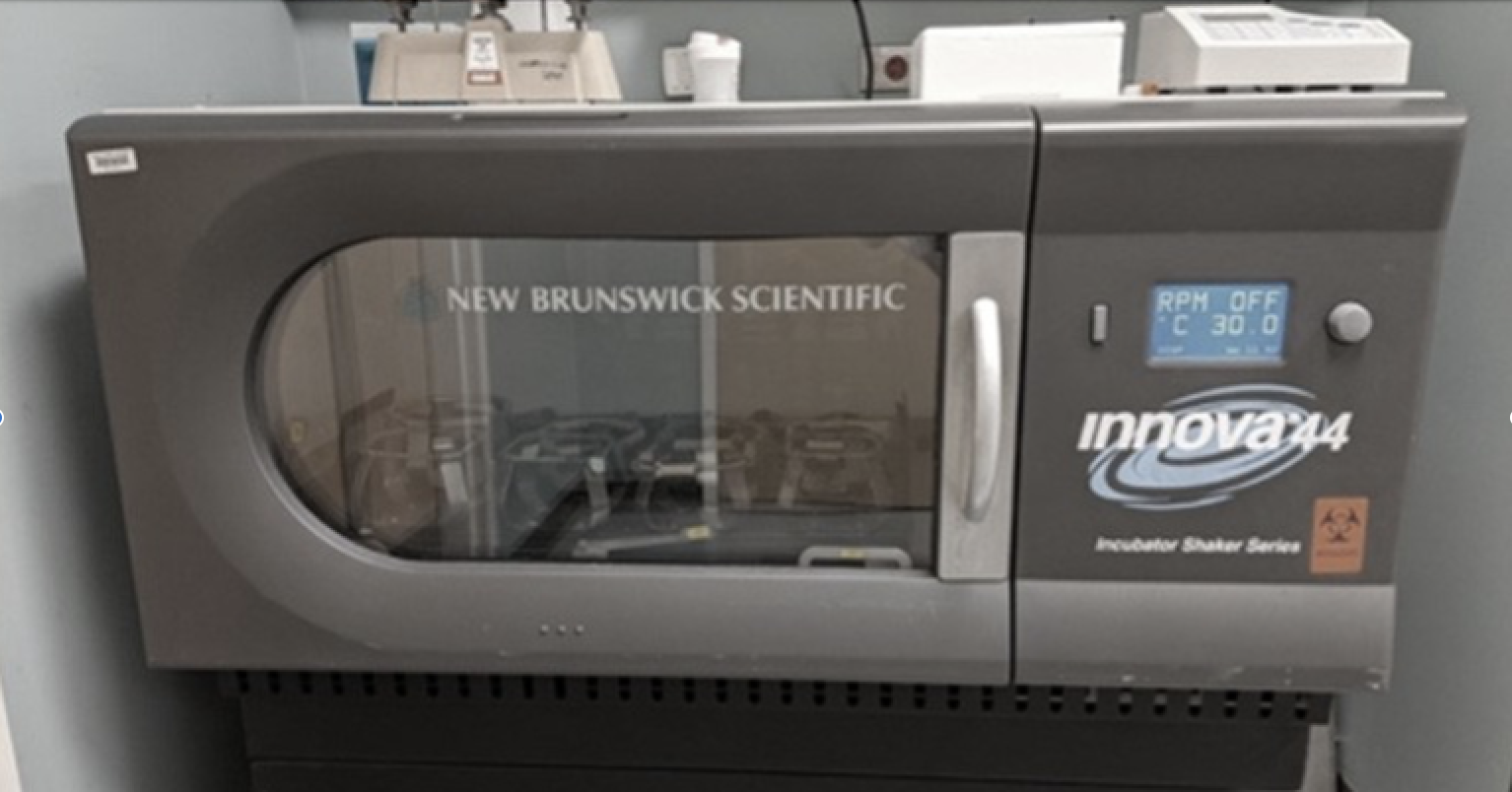
New Brunswick Innova 44R
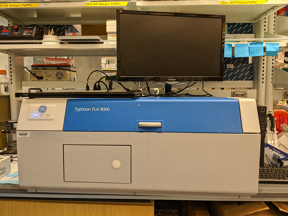
Typhoon FLA 9000
The Sarafianos lab has a BioTek Synergy Neo2 high-throughput multimode plate reader for luminescence, fluorescence, and absorbance assays. The Sarafianos lab also has both a ThermoFisher Scientific QuantStudio 3 Real-Time PCR System and a Thermo Scientific PikoReal Real-Time PCR System for qPCR analyses and thermal shift assays to screen for ligands/proteins that bind target proteins. Protein melting curves are analyzed using the associated QuantStudio Design and Analysis or PikoReal software package. The Sarafianos lab also has a Typhoon FLA 9000, which is a versatile laser scanner used to image biochemistry gels, and a New Brunswick Innova 44R refrigerated shaking incubator for bacteria induction and protein expression.
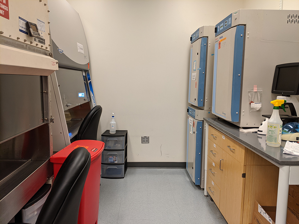
BSL2 cell culture room
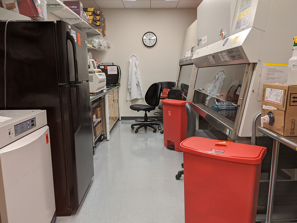
BSL2+ suite
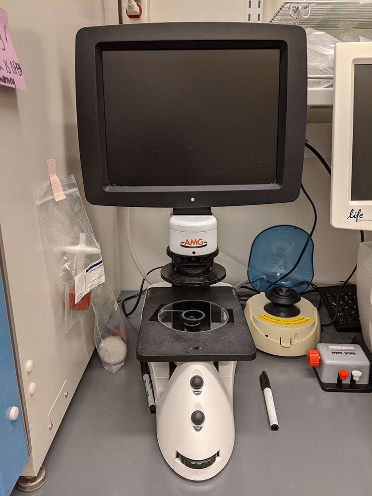
EVOS XL Core
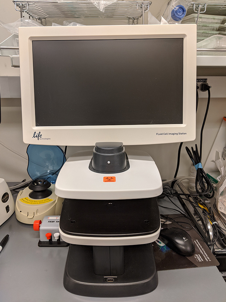
EVOS FLoid
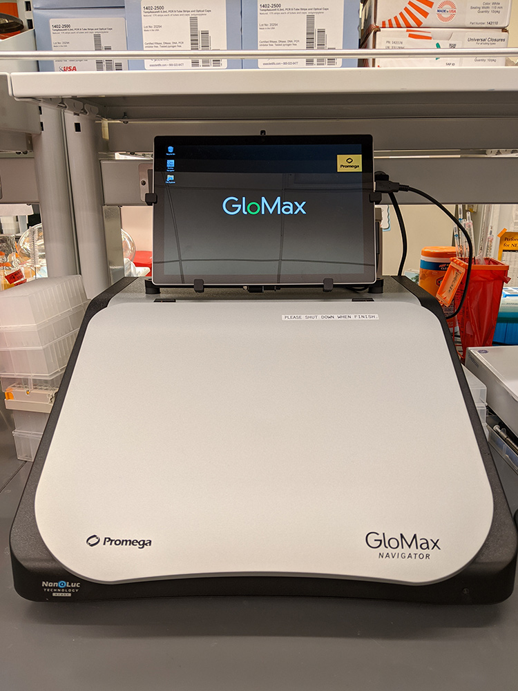
GloMax luminometer
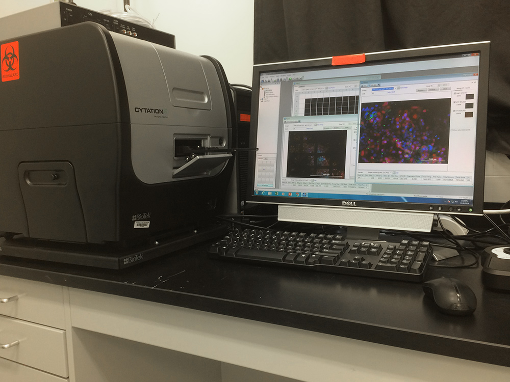
BioTek Cytation5
For protein chemistry and general molecular biology work:
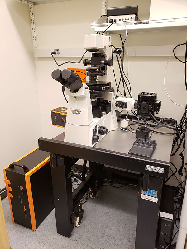
For high-resolution imaging, the Sarafianos lab has a Nikon C2 confocal system with 3-channel detector, LUN4 laser unit (405, 488, 561, and 640 nm solid-state lasers) on a TiS microscope equipped with epifluorescence illumination, 10x, 20x, and 60x objectives, and a manual stage. NIS-Elements C software is used to perform image analysis. For super-resolution imaging, the Sarafianos lab houses the HIVE Center’s Aberrior STEDYCON confocal and multicolor STED microscope on a Nikon TiS epifluorescence microscope with 10x, 20x, 40x, 60, and 100x objectives and a motorized stage. STED and confocal optics include 4 excitation laser lines (405, 488, 561, and 640 nm, all pulsed and all aligned by design; maintenance-free optical beam path), 775 nm STED laser, superior STED resolution <40 nm (typically 30 nm lateral, depending on dyes and objectives used). QUAD-beam scanner includes patent-protected scanning technology, scan lens-free design for minimal wavefront distortions, field of view up to 90 µm x 80 µm (with 100x objective), up to 1.1 frames/sec @ 512 x 512 pixels (line frequency up to 800 Hz), and quasi-simultaneous line-interleaved scanning, also between STED and confocal. Detection includes two STED and four confocal imaging channels, three single-photon-counting APD modules (extremely low dark counts to render the finest details and superior photon detection sensitivity (>62%), detection bands of 500-550 nm, 580-630 nm, and 650-700 nm (line multiplexes into four channels), DyanmicPlus detection, highly accurate time-gating for confocal and STED, and a motorized pinhole with 12 pinholes (10 µm to 200 µm). The system is complete with Aberrior’s image analysis software.
Nikon C2 confocal
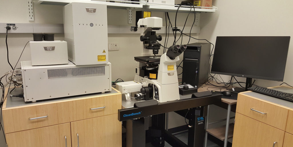
abberior STEDYCON
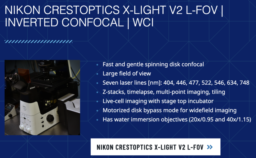
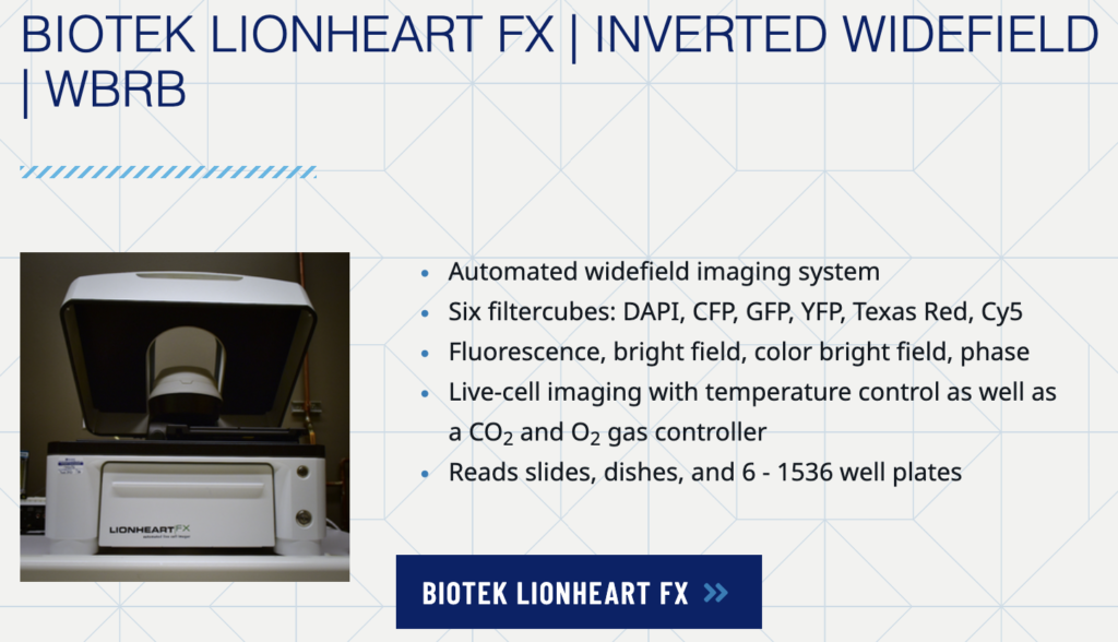
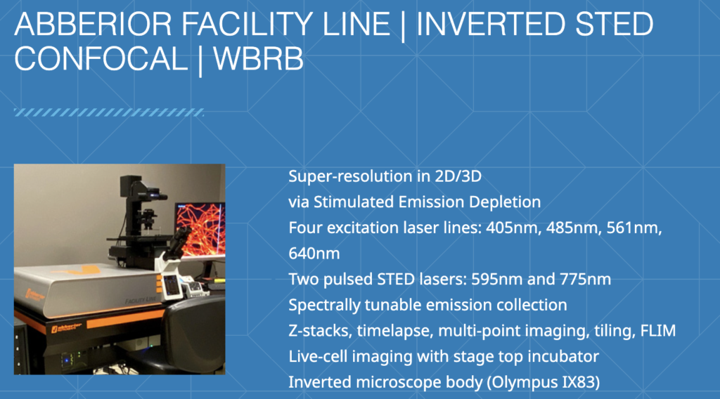
The Sarafianos Lab also has access to the Integrated Cellular Imaging core facility in our building for using a Olympus FV1000 inverted confocal microscope, a Nikon TE2000 Spinning Disk microscope for live cell imaging, and DeltaVision OMX Blaze structured illumination microscope for super-resolution 2D and 3D live cell imaging, providing an 8-fold increase in volume resolution compared to confocal microscopy. Additionally, the core contains workstations with a wide variety of image analysis packages (e.g. Imaris, FIJI, Volocity, MATLAB, Huygens, Cell Profiler, etc.) and expert staff to aid in analyses. The Integrated Cellular Imaging core also provides access to equipment across campus, including additional confocal and super-resolution microscopes, with capabilities including live-cell, multiphoton and TIRF imaging.
Nikon CrestOptics
BioTek Lionheart
abberior FACILITY line
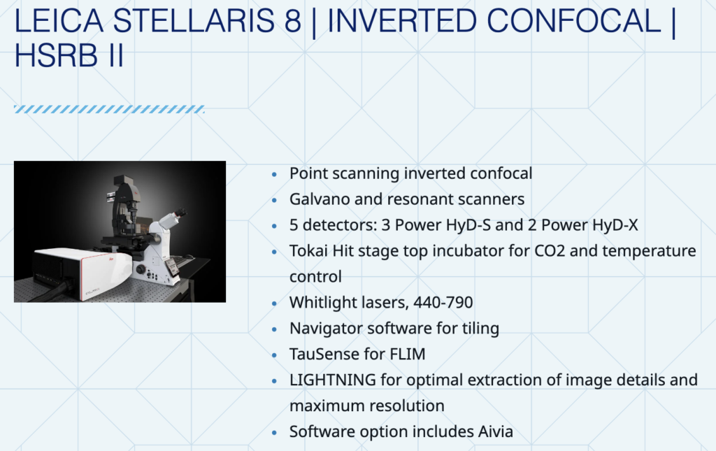
Leica STELLARIS 8
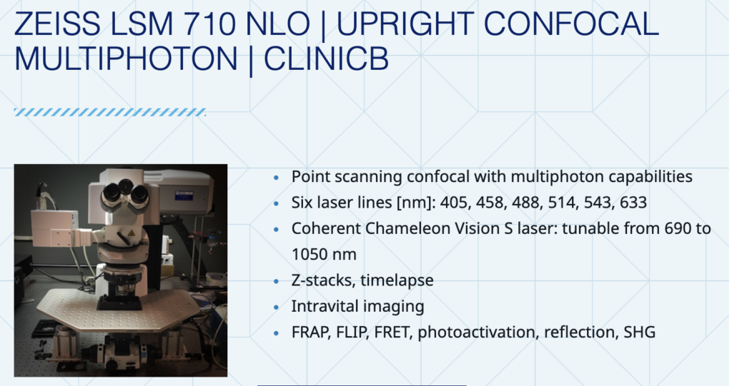
Zeiss LSM 710 NLO
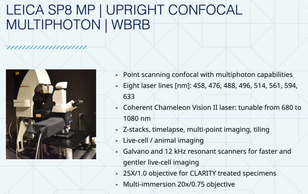
Leica SP8 MP
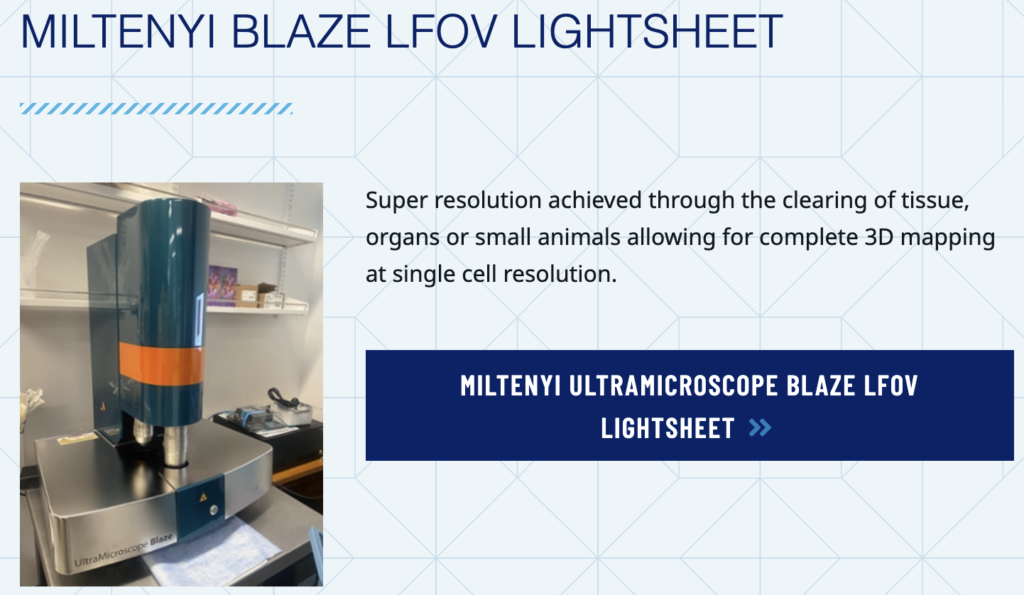
UltraMicroscope Blaze
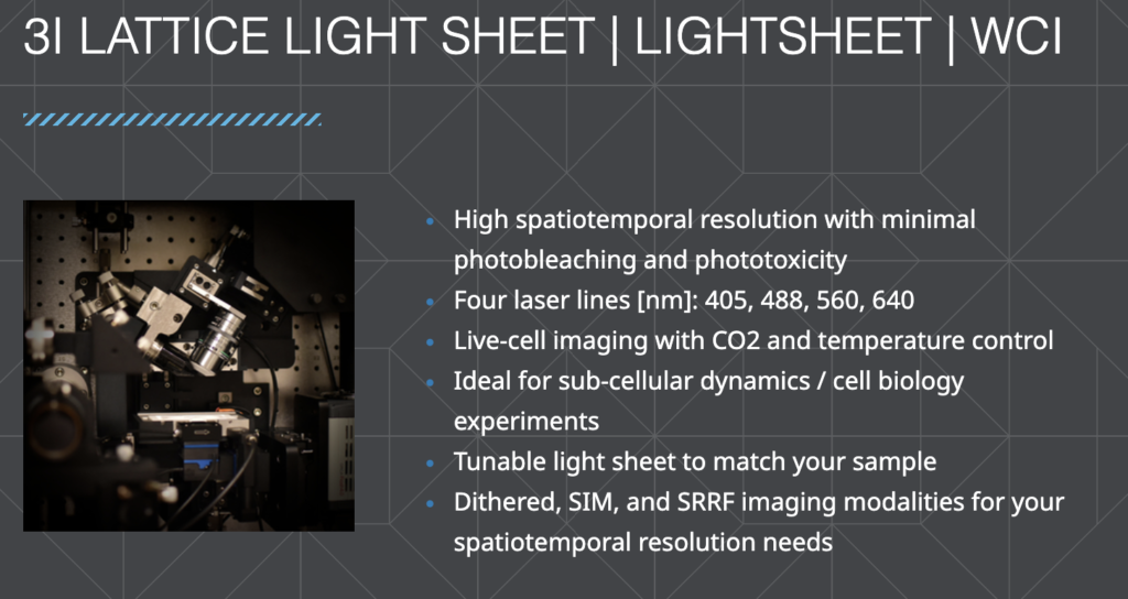
3i Lattice LightSheet
For electron microscopy experiments:
The Sarafianos lab has access to the an Integrated Electron Microscopy Core, which houses the equipment below.
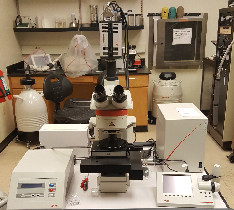
Leica EM Cryo CLEM
For cryo Correlative Light and Electron Microscopy (cryo-CLEM) experiments, the Sarafianos and Melikian labs jointly own a Leica EM Cryo CLEM microscope.The Leica EM Cryo CLEM is a dedicated cryo fluorescence microscope designed to maintain optimal cryo conditions for smooth, contamination-free sample handling throughout the CLEM workflow. The microscope provides high resolution imaging, fast image acquisition, and the unique software provides a seamless transfer of sample coordinates to different EM softwares for correlation of fluorescence and electron microscopy images.
For Transmission Electron Microscopy (TEM), there is a JEOL JEM-1400 120 kV LaB6 TEM with Gatan CCD cameras. Capable of all modes of TEM, including tomography of sectioned materials. This TEM has accelerating voltages of 40 kV – 120 kV, a LaB6 filament, Gatan CCD cameras, negative sheet film, image orientation system, motorized goniometer allows +/- 80° sample tilting, minimum Dose System (MDS) allows for imaging of beam-sensitive samples, SerialEM for semi-automated data acquisition, and beam blocker for electron diffraction work. Additionally, there is a Hitachi HT-7700 120 kV W (Tungsten) TEM with AMT CCD camera. This microscope is capable of all modes of TEM, including tomography and electron diffraction, has accelerating voltages of 40 kV – 120 kV, a W (Tungsten) filament, AMT CCD camera, image orientation system, motorized goniometer allows +/- 70° sample tilting, a Low Dose System that allows for imaging of beam-sensitive samples, SerialEM for semi-automated data acquisition, and a beam blocker for electron diffraction work. There is also an FEI Talos 120 kV TEM with an LaB6 filament and a 4k Ceta detector.
For cryo-Electron Microscopy (cryo-EM) there are two Gatan CP3s and two FEI Vitrobot Mark IVs for plunge freezing sample grids into liquid ethane. There is also a BAL-TEC HPM 010 High Pressure Freezing machine for preparing frozen specimens including thick samples and monolayer cell cultures
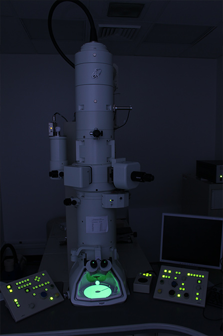
JEOL 1400 TEM
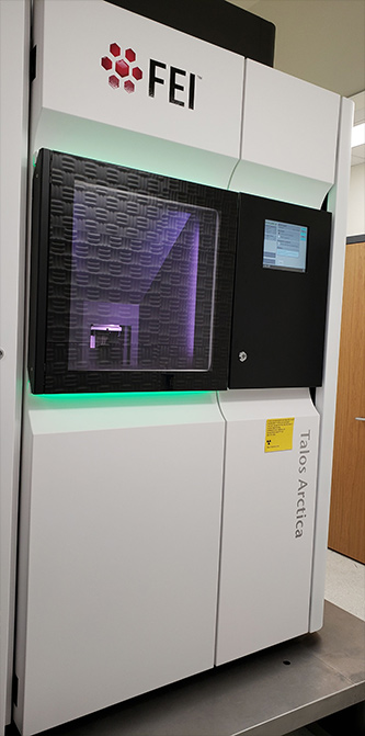
FEI Talos Arctica
For high-resolution imaging, there is an FEI Talos Arctica 200 kV TEM with BioQuantum/K2 direct electron detector (K3 upgrade) with Autoloader system, which is used primarily for acquisition of single-particle cryo-EM data.
There is also a JEOL JEM-2200FS 200 kV Field Emission TEM with an in-column Omega filter, Gatan US1000 CCD camera, Direct Electron Detector (DE-20) for high resolution imaging, motorized goniometer for +/- 80° sample tilting, minimum dose system for imaging of beam-sensitive samples, and SerialEM for semi-automated data acquisition, which is primarily used for acquisition of cryo-electron tomography data.
For Scanning Electron Microscopy (SEM) we have access to a Topcon DS-130F Field Emission SEM/STEM capable of in-lens and below-lens conventional SEM, and in-lens cryo-HRSEM. There is also a Topcon DS-150F Field Emission SEM /STEM with BSE (back-scattered electron detection) and capable of in-lens and below-lens conventional SEM, and in-lens cryo-HRSEM. This instrument has accelerating voltages of 0.5 kV – 30 kV, Schottky field emission sources, BSE and STEM systems, a Gatan CT-3500 cold stage, and an in-lens and below-lens imaging system.
For Sample Preparation, there is access to a Denton DV-602 Turbo Magnetron Sputter system with a chromium target, an EMSCOPE SC-500 Magnetron Sputter system with a gold target, a Denton Benchtop Turbo Carbon Evaporator, a Pelco Easy Glow glow discharger, Gatan 655 dry pumping station for cryo holders, a Leica UC6rt and UltracutS Ultramicrotome, a RMC PT-XL Ultramicrotome, Leica AFS cryo-substitution apparatus, and Leica UC6i/FC6 Cryo-Ultramicrotome.
For Imaging Data Processing and Analysis, there is access to three iMac workstations with Adobe, ImageJ, and IMOD software packages. There is also a Glacier computation cluster with 2 GPU nodes and 4 CPU nodes with image processing softwares including Relion, CisTEM, EMAN2/SPHIRE for processing of cryo-EM data.
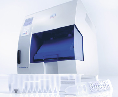
Qlagen EZ1
Additionally, the Sarafianos lab also has access to the general laboratory common resources in the Laboratory of Biochemical Pharmacology (LOBP), including fume hoods, biological safety cabinets, multiple refrigerators, -20 °C freezers and ultra-low freezers (-70 °C & -86 °C), a walk-in cold room, low speed and refrigerated centrifuges, analytical balances, autoclaves, incubators, UV spectrophotometers, a laser densitometer, Beckman ultracentrifuges, microtomes, lyophilizers, immuno-electrophoresis equipment, and gamma and liquid scintillation counters.
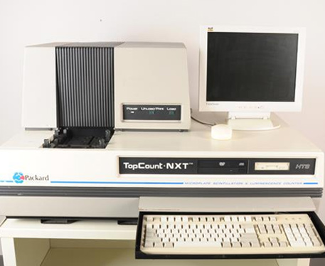
Qlagen EZ1
BSL-2* Laboratory: The Sarafianos lab has access to Dr. Raymond Schinazi’s BSL-2* facility at the LOBP, which is equipped with eight 6-foot laminar flow hoods (Nuaire), a Perkin Elmer TopCount NXT Microplate Scintillation and Luminescence Counter, a Filtermate Harvester, a Beckman Vi-cell XR cell viability analyzer and a Qiagen EZ1 Advanced XL DNA and RNA extractor. It also houses eight CO2 incubators, water baths, microscopes (Nikon, Olympus, EVOS), a refrigerator and ultra-low freezer, low-speed and refrigerated centrifuges, and two autoclaves.
The molecular biology & virology lab at LOBP is equipped with laminar flow hoods, microscopes, CO2 incubators, water baths, a cell harvester, an incubator shaker, a PerkinElmer JANUS robotic multi-channel pipetting system, an Epoch Biotek enzyme linked immunosorbent assay reader interfaced to a computer and printer, a Labcono centriVap complete, Beckman Avati J- 26S XPI centrifuge, Beckman XE-90 ultracentrifge, NewBrunswick shaking incubator, a MacsQuant flow cytometers/cell sorter (Miltenyi Biotec), and an Automacs Pro cell sorter (Miltenyi Biotec).
In addition, molecular biology laboratories house a Roche RT-PCR LightCycler 480 machine, an Applied Biosystems Prism RT-PCR machine, a MagnaPure DNA and RNA extractor, a Glomax multi detection system (Promega), a Gel Doc XR Imaging System, Western blot and sequencing apparatus, two BioRad 96- well thermal cyclers, three additional PCR thermal cyclers, a Pharos FX Plus Molecular Imager and Screen Eraser K (BioRad), and a GS Junior 454 Instrument for DNA sequencing system (Promega), a Gel Doc XR Imaging System, Western blot and sequencing apparatus, two BioRad 96- well thermal cyclers, three additional PCR thermal cyclers, a Pharos FX Plus Molecular Imager and Screen Eraser K (BioRad), and a GS Junior 454 Instrument for DNA sequencing.

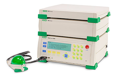
The Sarafianos lab also has access to a General Equipment Core that includes the Beckman Optima XPN-100 Ultracentrifuge, Beckman Avanti J-26S XPI Centrifuge (both centrifuges are housed within the Sarafianos lab space), and Bio-Rad Gene Pulser Xcell Electroporation System.
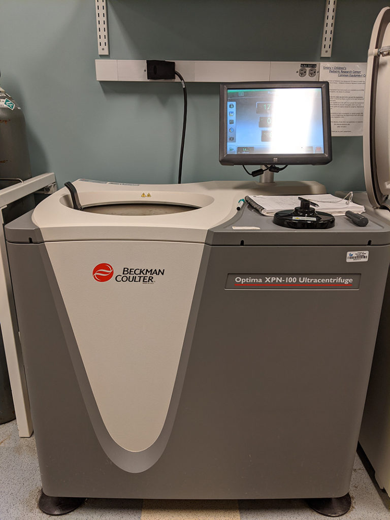
Optima-XPN-100-Ultracentrifuge
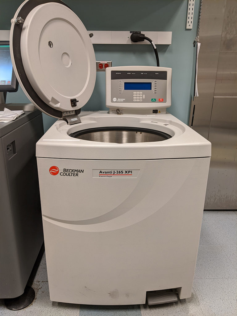
Avanti-J-26S-XPI-Centrifuge
